
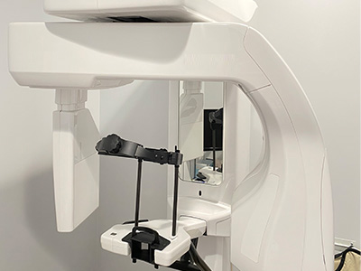
CBCT

At the office of Ashford Dental Sherman Oaks, we rely on advanced diagnostic tools to deliver precise, predictable dental care. One of the most important technologies in our imaging suite is cone-beam computed tomography (CBCT), which produces three-dimensional views of the teeth, jaws, and surrounding structures. These detailed images help our clinicians make better-informed decisions and tailor treatment plans to each patient’s unique anatomy.

Our practice uses a modern CBCT system designed to capture high-resolution scans with targeted exposure. The result is clear, clinically useful data that supports a wide range of services — from implant planning and endodontics to evaluation of airway and temporomandibular joint concerns. We prioritize diagnostic accuracy while keeping patient comfort and safety at the forefront of every exam.

How CBCT differs from traditional dental X-rays

Conventional dental X-rays create two-dimensional images that can obscure important anatomical relationships. CBCT, by contrast, acquires volumetric data, allowing clinicians to view structures from multiple angles and in cross-section. This three-dimensional perspective reveals depth, bone thickness, root canal anatomies, and spatial relationships that are otherwise hidden on flat films.

The ability to scroll through thin slices of a scan lets dentists inspect anatomy layer by layer. For example, a cross-sectional view can show the exact width of the jawbone where an implant is planned, or the proximity of a tooth root to a sinus cavity. That level of detail leads to more predictable treatment planning and fewer surprises during procedures.

Technically, CBCT uses a cone-shaped X-ray beam and a digital detector to reconstruct a 3D volume in a single rotation. Its design balances image quality and efficiency, capturing the information needed for dental diagnosis without the time and complexity of medical CT scans. The result is a practical, chairside imaging option that integrates well into modern dental workflows.

Clinical uses where CBCT makes a measurable difference

CBCT is invaluable for dental implant planning because it shows both the height and width of bone, the location of vital structures like nerves and sinuses, and the relationship between neighboring teeth. That spatial information supports precise implant placement and the selection of appropriately sized fixtures, helping to minimize complications and support long-term success.

In endodontics, CBCT reveals complex root canal configurations, periapical pathology, and fractures that might not be visible on standard films. Clinicians can identify hidden canals or subtle lesions and tailor root canal procedures with greater confidence. This often improves diagnosis for persistent symptoms and supports more conservative, tooth-preserving care.

Beyond implants and endodontics, CBCT aids in assessing impacted teeth, airway dimensions for sleep-breathing evaluations, and temporomandibular joint (TMJ) disorders. It also assists in oral surgery planning, orthodontic assessment, and evaluation of facial trauma or pathology, making it a versatile diagnostic tool across many dental specialties.

What patients can expect during a CBCT visit

A CBCT scan is quick and noninvasive. The patient typically stands or sits while the scanner rotates around the head, and a full-volume scan can be completed in less than a minute. Because the device is designed for dental imaging, the process is usually more comfortable and less intimidating than medical imaging for most people.

Before the scan, clinical staff will explain the process and position the patient to ensure accurate results. We use stabilization aids when needed to minimize movement and capture sharp images. The short scan time reduces the likelihood of motion artifacts, which helps ensure that the images are clinically useful on the first attempt.

After acquisition, the digital volume is reviewed by the treating clinician. Cross-sectional views and 3D renderings are used to identify key findings and to plan next steps. Because CBCT images are stored digitally, they can be shared with specialists or incorporated into digital treatment workflows, facilitating collaborative care when necessary.

Safety, radiation considerations, and quality control

Radiation safety is an important consideration for any imaging modality. Modern dental CBCT units are engineered to provide the lowest radiation dose practical for the diagnostic task at hand. Clinicians select field-of-view (FOV) and exposure settings that capture only the anatomy needed, following the principle of ALARA — as low as reasonably achievable.

Our office follows industry guidelines and manufacturer recommendations for routine calibration and quality assurance checks to maintain image consistency and minimize unnecessary exposure. CBCT is used judiciously, reserved for clinical situations where the additional information will directly affect diagnosis or treatment planning.

For patients who are pregnant, very young, or have specific sensitivities, clinicians evaluate the risk-benefit balance before recommending any radiographic exam. When CBCT is warranted, protective measures and appropriate shielding are employed to reduce exposure to non-target areas.

How CBCT integrates with digital treatment planning

CBCT data can be combined with intraoral scans, digital models, and planning software to create a complete virtual treatment environment. For implant therapy, this digital integration allows the design of guided surgical templates that translate the virtual plan into precise clinical placement. The result is smoother procedures and enhanced predictability.

When multiple specialists are involved — for example, a surgeon, restorative dentist, and lab technician — CBCT volumes provide a common reference point for communication. Digital files can be exported in standard formats and reviewed collaboratively, improving coordination and reducing misinterpretation that can arise from two-dimensional images alone.

Even when advanced guides are not required, CBCT contributes to better-informed clinical decisions. By visualizing anatomy in three dimensions, dentists can choose approaches that preserve healthy structure, avoid critical landmarks, and anticipate technical challenges before a procedure begins.

In summary, CBCT is a powerful diagnostic tool that enhances clinical insight across many areas of dentistry. At Ashford Dental Sherman Oaks, we use CBCT thoughtfully to improve treatment planning, reduce uncertainty, and deliver care that is both precise and patient-centered. If you have questions about whether CBCT is appropriate for your situation, please contact us to learn more.

Frequently Asked Questions

What is CBCT and how does it work?

+

Cone-beam computed tomography (CBCT) is a three-dimensional dental imaging technique that captures volumetric data of the teeth, jaws and surrounding structures in a single rotation. The scanner projects a cone-shaped X-ray beam onto a digital detector and reconstruction software compiles the acquired views into a detailed 3D volume. This volumetric data can be reformatted into cross-sectional slices, panoramic views and 3D renderings to reveal spatial relationships that two-dimensional films cannot show.

Because CBCT provides depth information, clinicians can evaluate bone thickness, root trajectories and anatomical landmarks with greater precision. The speed and convenience of modern dental CBCT units make them practical for chairside use, delivering clinically relevant images that support diagnosis and treatment planning across multiple dental specialties.

How does CBCT differ from conventional dental X-rays?

+

Conventional dental X-rays produce two-dimensional images that compress depth information into a flat plane, which can obscure overlapping structures and hide critical anatomical detail. CBCT acquires volumetric data so clinicians can view anatomy from multiple angles and scroll through thin cross-sectional slices to inspect depth and relationships. This three-dimensional perspective is particularly valuable when assessing bone volume, nerve position and the spatial orientation of roots or impacted teeth.

While periapical and bitewing films remain useful for routine screening and detecting caries, CBCT is selected when additional spatial information is needed to answer specific clinical questions. The choice between 2D and 3D imaging is guided by the diagnostic task, with CBCT reserved for situations where the additional information will change treatment decisions.

What clinical situations commonly require CBCT imaging?

+

CBCT is commonly indicated for dental implant planning, assessment of complex endodontic anatomy, evaluation of impacted or supernumerary teeth, and investigation of facial trauma or pathology. It is also valuable for TMJ analysis, airway assessment in sleep-breathing evaluations, and pre-surgical planning for extractions or orthognathic procedures. Each of these applications benefits from the volumetric insights that reveal bone volume, canal morphology and the relationship of dental structures to vital anatomy.

Clinicians select CBCT when conventional imaging does not provide sufficient information to plan safely and predictably. The decision is based on the clinical history, examination findings and the likelihood that 3D data will materially affect diagnosis or the treatment approach.

How does CBCT improve implant planning and placement?

+

CBCT allows precise measurement of bone height, width and density, and shows the proximity of implants to critical structures such as the inferior alveolar nerve and maxillary sinuses. With these measurements clinicians can select optimal implant length, diameter and position to maximize stability and avoid anatomical complications. Three-dimensional visualization reduces intraoperative surprises and supports predictable outcomes by informing both the restorative plan and the surgical approach.

When combined with digital planning software, CBCT volumes can be used to design guided surgical templates that transfer the virtual plan to the clinical setting. This digital integration improves accuracy during placement, facilitates prosthetic-driven planning and enhances communication between surgeons, restorative dentists and dental laboratories.

Can CBCT help with endodontic diagnosis and treatment?

+

Yes. CBCT is especially useful in endodontics for revealing complex root canal anatomy, identifying previously undetected canals, and detecting vertical root fractures or periapical pathology that may be masked on 2D films. The ability to view teeth in cross-section helps clinicians locate accessory canals and assess the extent and location of lesions with greater confidence. This information can change the treatment plan and support more conservative, tooth-preserving care.

CBCT also aids in evaluating persistent symptoms after prior treatment by clarifying whether retained infection, missed anatomy or a structural defect is present. When used judiciously and interpreted by trained clinicians, CBCT improves diagnostic accuracy and can guide more targeted endodontic therapy.

What should patients expect during a CBCT appointment at Ashford Dental Sherman Oaks?

+

A CBCT scan is a quick, noninvasive procedure that typically takes less than a minute of actual exposure time, although patient positioning and setup may take a few additional minutes. The patient usually stands or sits while the machine rotates around the head, and staff use positioning aids and instructions to minimize movement. Most people find the experience comfortable and less confining than many medical imaging environments.

After the scan the clinician reviews cross-sectional images and 3D renderings to identify findings and plan next steps. Because the images are digital, they can be shared with specialists or incorporated into digital treatment workflows to support coordinated care and clear communication among the treatment team.

Is CBCT safe and what are the considerations for radiation exposure?

+

Modern dental CBCT units are engineered to deliver the lowest radiation dose consistent with obtaining diagnostic-quality images, and clinicians follow the ALARA principle — as low as reasonably achievable — when selecting imaging parameters. Field-of-view (FOV) selection, voxel size and exposure settings are tailored to capture only the anatomy necessary for the clinical question, which minimizes unnecessary exposure. Compared with medical CT scans, dental CBCT typically uses substantially lower doses because it is focused on a limited region and optimized for dental applications.

Special precautions are taken for pregnant patients, very young children or those with specific sensitivities, and clinicians weigh the diagnostic benefit against any potential risk before recommending imaging. When a CBCT exam is indicated, appropriate shielding and dose-minimizing protocols are used to protect non-target tissues and ensure patient safety.

How is CBCT data integrated with other digital tools in treatment planning?

+

CBCT volumes are often merged with intraoral scans, digital models and planning software to create a comprehensive virtual treatment environment. This fusion of data enables prosthetically driven implant planning, design of guided surgical templates and accurate simulation of restorative outcomes. The combined datasets improve visualization of spatial relationships between planned restorations and the underlying anatomy.

Shared digital files also facilitate collaboration among clinicians, specialists and dental laboratories by providing a common visual reference. Standard export formats allow CBCT data to be reviewed in multiple software platforms, supporting coordinated, multidisciplinary care and more predictable procedural execution.

What are the limitations of CBCT imaging that patients should understand?

+

Although CBCT provides excellent detail for hard tissues and bony anatomy, its soft-tissue contrast is limited compared with medical CT or MRI, so it is not a substitute when high-resolution soft-tissue imaging is required. Metal restorations and orthodontic appliances can produce artifacts that degrade image quality in regions of interest, and small structures can be affected by voxel size and user-selected parameters. Image interpretation also requires clinical correlation; CBCT findings must be evaluated alongside examination and history to avoid overreliance on imaging alone.

Because of these limitations, clinicians reserve CBCT for situations where the expected diagnostic benefit outweighs the modality’s constraints. Careful case selection and proper technique help maximize the value of CBCT while acknowledging what the technology can and cannot reveal.

How does the office of Ashford Dental Sherman Oaks ensure CBCT image quality and reliable interpretation?

+

The office maintains CBCT quality through routine calibration, manufacturer-recommended maintenance and regular performance checks to ensure consistent image fidelity. Clinicians and staff follow standardized imaging protocols, select appropriate field-of-view and exposure settings for each indication, and use stabilization techniques to minimize motion artifacts. Training in image acquisition and interpretation ensures that scans are both diagnostically useful and acquired with patient safety in mind.

Scans are reviewed on calibrated displays and incorporated into the patient's digital record to support treatment planning and interprofessional communication. When cases benefit from specialist input, CBCT data can be shared securely with consultants to support collaborative decision-making and optimize patient outcomes.

Hours of Operation

Monday
9:00 am - 6:00 pm
Tuesday
Closed
Wednesday
9:00 am - 6:00 pm
Thursday
9:00 am - 1:00 pm
Friday
8:30 am - 4:00 pm
Saturday
9:00 am - 2:00 pm
(Every Other)