
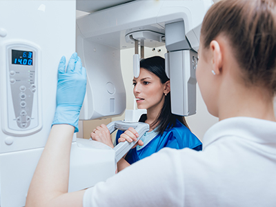
Digital radiography replaces traditional film with electronic sensors and advanced imaging software to capture dental X-rays. Instead of developing film in a darkroom, the sensor records the exposure and converts it into a digital file that becomes part of a patient’s chart. The result is a cleaner, faster workflow that supports more precise recordkeeping and better clinical decision-making.
For patients, the most tangible benefit is speed: images appear on-screen within seconds, allowing the dentist to review findings and explain concerns during the same appointment. For clinicians, immediate access to high-resolution images helps identify issues that might be missed on older film, supporting earlier intervention when needed. The technology is a foundational tool that enhances routine exams, restorative planning, and monitoring of long-term oral health.
Because the process captures images digitally, those files can be archived, annotated, and retrieved without loss of quality. Digital radiography also integrates smoothly with other chairside technologies—such as intraoral cameras and 3D imaging systems—allowing clinicians to combine visual information for a more complete diagnostic picture. That interoperability improves treatment planning and helps patients understand their options.
Contemporary sensors produce images with greater detail and contrast than traditional film, which helps clinicians spot small changes in tooth structure and surrounding bone. Software tools allow careful adjustment of brightness, contrast, and magnification so specific areas of concern can be examined closely without repeating exposures. These enhancements are especially valuable when assessing cavities between teeth, the health of previous restorations, or subtle changes in the jawbone.
There are several types of digital sensors used in dentistry, each designed to balance comfort, image quality, and durability. Sensors are thin and ergonomically shaped to fit comfortably in the mouth while capturing the necessary anatomy. Because images can be reviewed instantly, the clinician can confirm that the picture shows the intended area and retake it immediately if needed, rather than discovering an issue later when the film is developed.
Beyond single-image review, digital radiography facilitates comparison across appointments. Clinicians can overlay or place images side-by-side, making it easier to track healing, evaluate the progression of a lesion, or confirm the stability of a restoration. This temporal perspective strengthens clinical judgment and provides a clear visual record for ongoing care.
One of the most practical advantages of digital radiography is the ability to share images quickly and securely. Digital files can be transmitted to specialists, labs, or referring clinicians without physical transfer of films, which speeds consultations and reduces delays in care. When multiple providers need to collaborate—such as for implant planning or complex restorative work—everyone can see the same high-quality images simultaneously.
Instant access to images also enhances communication with patients. Dentists can display radiographs on the operatory monitor, point out areas of concern, and walk patients through recommended treatment steps. Visual explanations often help patients understand why a procedure is advised and what the expected outcome will be, supporting informed decision-making.
From a records standpoint, digital images are easier to organize and index, improving continuity of care. Secure storage within the electronic health record ensures images remain linked to the correct patient and appointment, and many systems include audit trails and access controls to maintain privacy and regulatory compliance.
Digital radiography typically requires less radiation exposure than conventional film-based X-rays because modern sensors are more sensitive to radiation. Lower exposure helps reduce cumulative dose over time, an important consideration for patients who require regular imaging as part of ongoing dental care. Clinicians still follow established safety protocols, but the technology itself contributes to minimizing exposure whenever possible.
Another practical benefit is the elimination of chemical processing. Traditional film development requires solutions and materials that must be handled, stored, and disposed of according to environmental regulations. Digital imaging avoids these chemicals entirely, reducing waste and simplifying infection-control procedures in the operatory and processing areas.
Because images are captured and stored electronically, there is also less physical material to manage—no film envelopes or storage boxes—making recordkeeping more compact and reliable. The combination of lower radiation and reduced environmental impact reflects how modern dental technologies can improve both patient safety and clinic efficiency.
Digital radiographs are an integral part of routine exams, treatment planning, and monitoring. During a comprehensive evaluation, images help reveal interproximal decay, evaluate the health of tooth roots and surrounding bone, and assess the fit and integrity of restorations. For restorative and surgical procedures, radiographs provide the anatomical roadmap clinicians use to plan precise, predictable care.
When complex cases require collaboration, digital images are shared with specialists to support coordinated treatment. Whether preparing for implant placement, evaluating endodontic needs, or assessing periodontal support, the availability of clear, immediate imagery improves clinical outcomes and helps patients feel involved in their care plan.
Throughout every visit, the practice prioritizes patient comfort and education. Clinicians take the time to explain findings using on-screen images, answering questions and outlining next steps. By combining clinical expertise with modern imaging tools, the office delivers diagnostic accuracy while keeping patients informed and involved in decisions about their oral health.
In summary, digital radiography is a fast, precise, and patient-friendly imaging method that enhances diagnosis, communication, and safety. At Ashford Dental Sherman Oaks we rely on these tools to support thorough exams and collaborative treatment planning. If you have questions about how digital imaging fits into your care, please contact us for more information.
Digital radiography uses electronic sensors and imaging software to capture dental X-rays and store them as digital files. These images appear on-screen within seconds, eliminating the need for film development and speeding clinical workflows. Because files are digital, they can be archived, annotated, and retrieved without degradation, which improves recordkeeping and continuity of care.
The technology matters because it enhances diagnostic accuracy and patient communication. High-resolution images and software tools allow clinicians to adjust contrast, magnification, and brightness to reveal subtle changes in teeth and bone. Faster access to reliable images supports earlier intervention and more predictable treatment planning.
Digital radiography replaces chemical film and darkroom processing with sensors that convert X-ray exposure into electronic images. The sensors are more sensitive to radiation and the images are available immediately, which reduces the need for retakes and shortens appointment times. Digital files also avoid the physical storage and chemical disposal associated with film, simplifying office logistics and environmental compliance.
Another key difference is image manipulation and integration. Digital images can be enhanced, measured, and compared side-by-side or overlaid to track changes over time, and they integrate with other digital tools such as intraoral cameras and electronic health records. This interoperability improves diagnostic confidence and facilitates collaboration among providers.
Digital dental X-rays generally require less radiation than conventional film X-rays because modern sensors are more efficient at detecting exposure. Dentists and staff follow established safety protocols, including proper shielding and techniques to minimize dose, and they take images only when clinically indicated. The goal is to balance diagnostic benefit with the lowest reasonable exposure for each patient.
For patients with specific concerns—such as pregnancy or a history of frequent imaging—clinicians discuss the rationale for any recommended radiographs and consider alternatives when appropriate. Overall, digital imaging contributes to dose reduction while maintaining or improving diagnostic quality, which supports safer long-term care.
During a comprehensive exam, digital radiographs reveal details that cannot be seen in a visual inspection alone, such as interproximal decay, root and bone health, and the integrity of restorations. Clinicians capture the images, review them immediately on-screen, and use software tools to highlight concerns and confirm that the intended anatomy is visible. This immediate feedback reduces the chance of missed views and allows the dentist to address findings during the same visit.
At Ashford Dental Sherman Oaks clinicians use digital images to document baseline conditions and to monitor changes over time. Radiographs also guide restorative and surgical planning by showing anatomical landmarks, bone levels, and the relationships between teeth and surrounding structures. Patients benefit from clear visual explanations that support informed decisions about their care.
The imaging process is quick and minimally invasive: a thin sensor is positioned in the mouth, and the X-ray unit produces a brief exposure while the patient remains still. Because the sensor is designed for comfort, most patients experience only mild pressure for a few seconds, and images appear on the monitor almost instantly. If a picture does not show the intended area, the clinician can retake it immediately to ensure diagnostic quality.
Clinicians explain the purpose of each image and review findings with the patient using on-screen displays. This visual review helps patients understand the reasons behind recommended treatments and allows the dentist to answer questions in real time. Staff also follow routine safety measures such as use of thyroid collars or other shielding when indicated.
Digital radiography provides higher-resolution images and software tools that enhance contrast, magnification, and measurement, making it easier to detect small cavities, monitor bone levels, and evaluate root structures. These capabilities reduce diagnostic uncertainty and help clinicians identify conditions at earlier stages when treatment is often simpler and more effective. Comparison tools also allow side-by-side review of images from different dates to assess progression or healing.
Integration with other digital technologies strengthens treatment planning by combining visual information into a cohesive view. For example, clinicians can correlate intraoral camera photos with radiographs or use radiographic measurements when planning restorations or implant placement. This comprehensive approach supports precise, predictable care and clear communication with specialists when collaboration is needed.
Digital radiographs can be transmitted electronically to specialists, labs, or referring clinicians, which speeds collaboration and reduces delays in care. Transfers are typically performed through secure channels within the electronic health record or via encrypted file exchange to maintain data integrity. Sharing digital images eliminates the need for physical films and allows multiple providers to view the same high-quality images simultaneously.
Privacy protections include access controls, audit trails, and encryption used by modern dental record systems to ensure that images remain linked to the correct patient and are accessed only by authorized personnel. Patients can discuss any concerns about data sharing with their clinician, who will explain how images are transmitted and stored in compliance with applicable privacy regulations.
Frequency of imaging is individualized based on a patient’s oral health, risk factors, and treatment needs rather than a fixed schedule for everyone. Patients with active disease, a history of frequent decay, or ongoing restorative or periodontal treatment may need images more often, while low-risk patients may require them less frequently. Dentists follow evidence-based guidelines and clinical judgment to recommend imaging at appropriate intervals.
During routine visits clinicians review symptoms, clinical exam findings, and the patient’s dental history to determine whether new radiographs are warranted. This tailored approach ensures that imaging provides clinical value while minimizing unnecessary exposure. Patients are encouraged to ask about the reason for any recommended images and how the results will influence their care plan.
Three-dimensional cone-beam computed tomography (CBCT) is preferred when detailed spatial information about teeth, bone, and anatomical structures is required for diagnosis or complex treatment planning. Indications include implant placement, evaluation of impacted teeth, assessment of jaw pathology, and certain endodontic or surgical cases where two-dimensional images do not provide sufficient detail. CBCT produces volumetric data that reveal relationships and depths not visible on standard radiographs.
Because CBCT delivers a higher radiation dose than routine intraoral digital images, clinicians reserve its use for cases where the additional information will change the treatment approach. The decision to obtain CBCT is based on clinical need and is made after discussing benefits, alternatives, and any patient-specific considerations. When CBCT is used, staff follow established protocols to limit field of view and exposure to what is necessary for the diagnostic task.
The practice combines digital radiographs with intraoral cameras, electronic health records, and three-dimensional imaging to create a comprehensive diagnostic workflow. Images are reviewed alongside clinical photos and patient histories to form a clear clinical picture, which improves treatment planning and patient education. This integrated approach enables clinicians to correlate findings from multiple modalities and make well-supported recommendations.
Integration also streamlines coordination with specialists and laboratories by providing standardized, high-quality images for collaboration. Digital records reduce duplication, improve tracking of outcomes over time, and support efficient clinical communication. Patients benefit from clearer explanations and more predictable care when multiple technologies work together in the diagnostic process.
Phone: